Please wait while we open this resource.


K

Clinical reviews
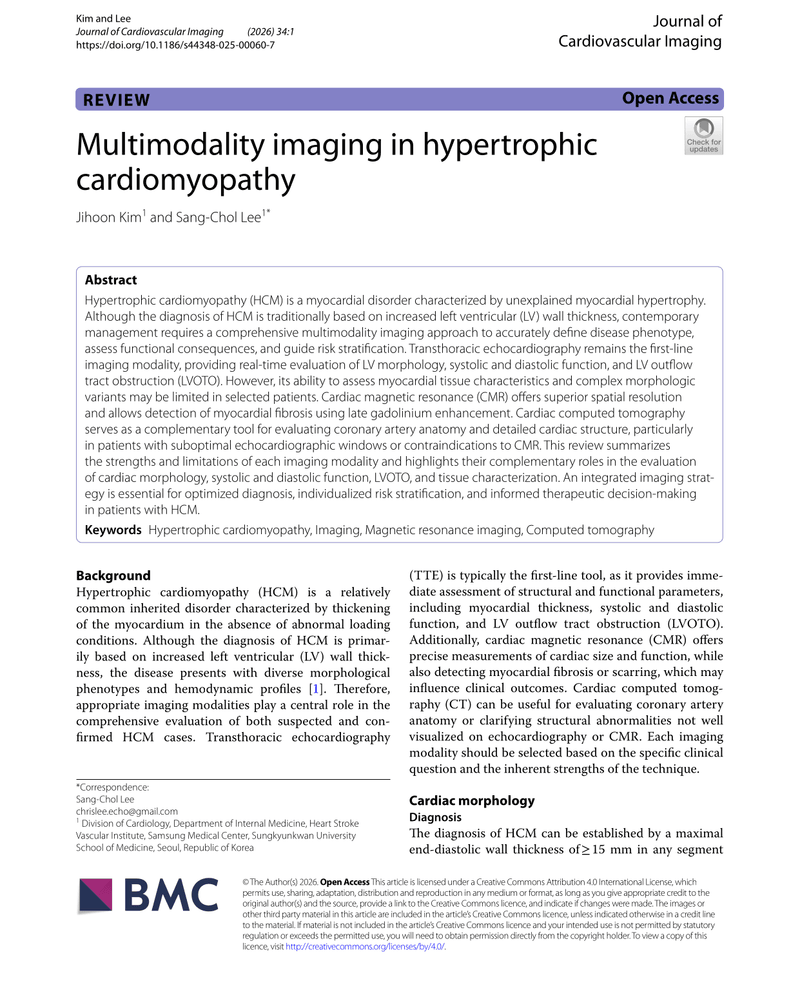
MULTIMODALITY IMAGING IN HYPERTROPHIC CARDIOMYOPATHY
Jihoon Kim and Sang-Chol Lee
PDF10 pages2.5 MB
Hypertrophic cardiomyopathy (HCM) is characterized by unexplained myocardial hypertrophy, and its diagnosis traditionally relies on increased left ventricular wall thickness. However, contemporary management necessitates a comprehensive imaging strategy to accurately define disease phenotype, assess functional consequences, and guide risk stratification. This review summarizes the roles of transthoracic echocardiography, cardiac magnetic resonance, and cardiac computed tomography in evaluating cardiac morphology and function, emphasizing the need for an integrated approach to optimize diagnosis and therapeutic decision-making in HCM patients.
Sign in to open
Ask this resource
Answers use this document only.
Your first question is free.
More like this
Resources from the same specialty and type. Curated links will replace these when added.
Clinical Resources
Documents that extend or complement this resource.
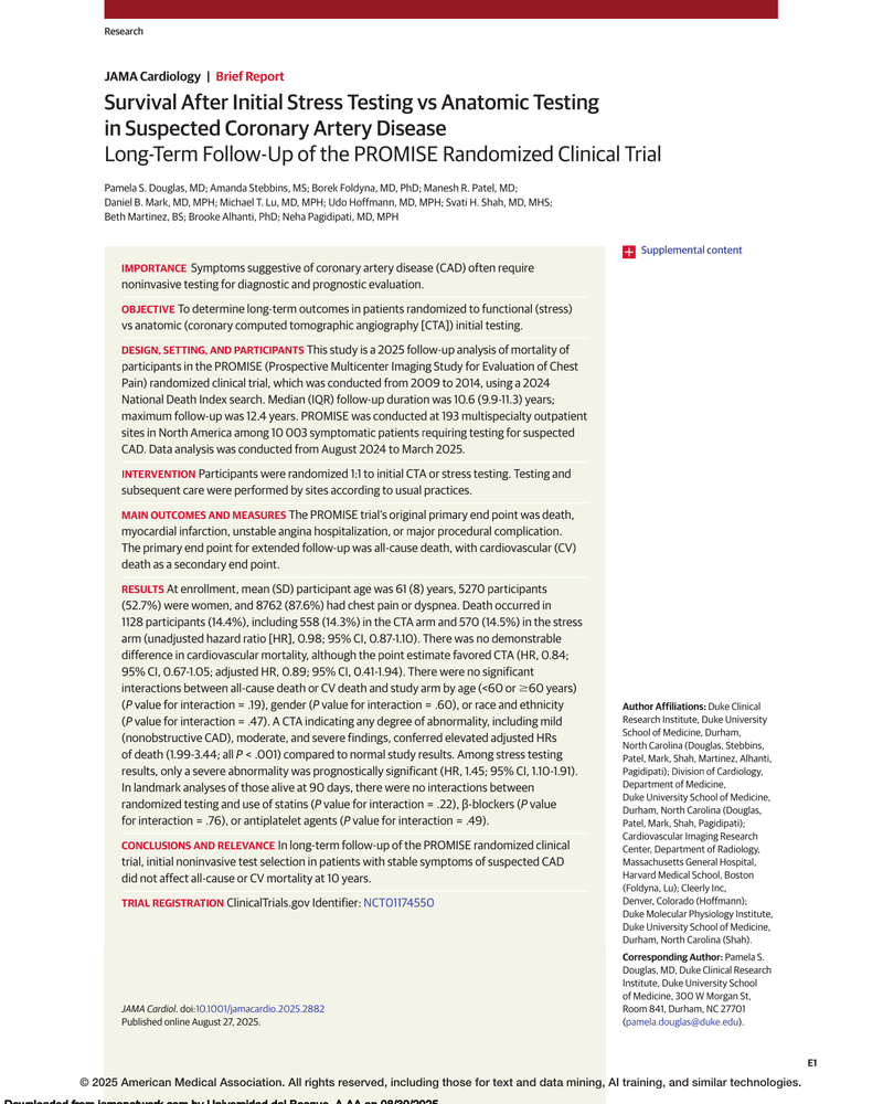
SURVIVAL AFTER INITIAL STRESS TESTING VS ANATOMIC TESTING IN SUSPECTED CORONARY ARTERY DISEASE
Same clinical topic · Cardiology

PRACTICAL CARDIO-ONCOLOGY
Same clinical topic · Cardiology

OXFORD CARDIOLOGY LIBRARY: Hypertension
Same clinical topic · Cardiology

CARDIAC INVOLVEMENT IN PARASITIC INFECTIONS
Same clinical topic · Cardiology
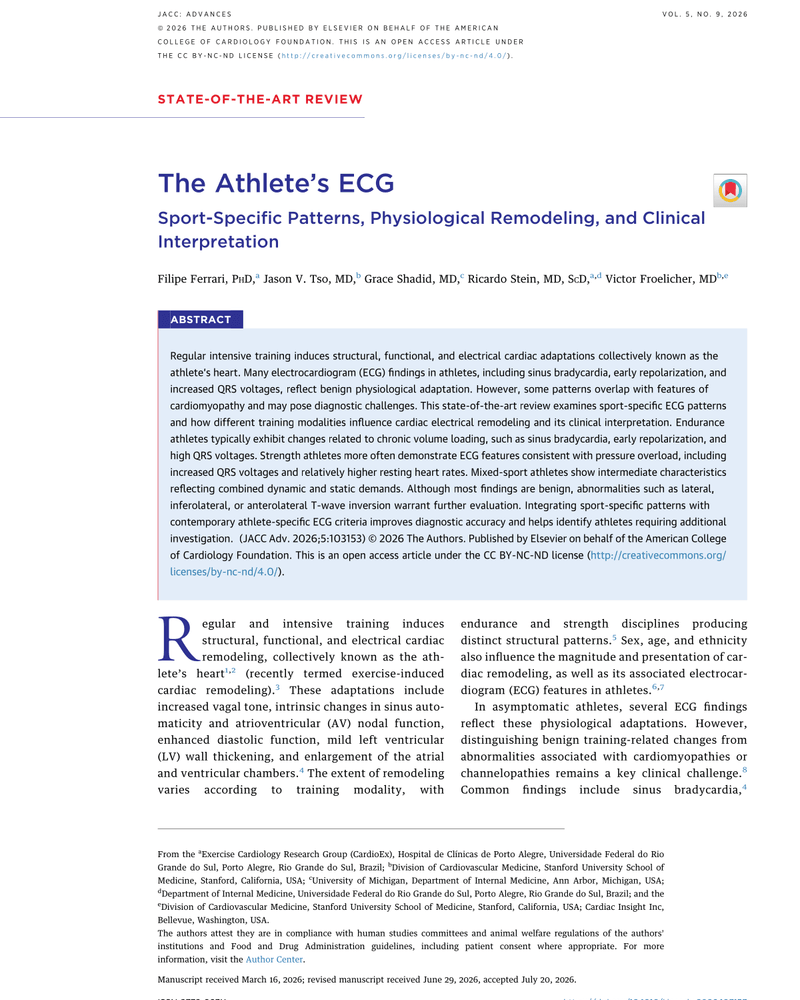
The Athlete’s ECG
Same clinical topic · Cardiology
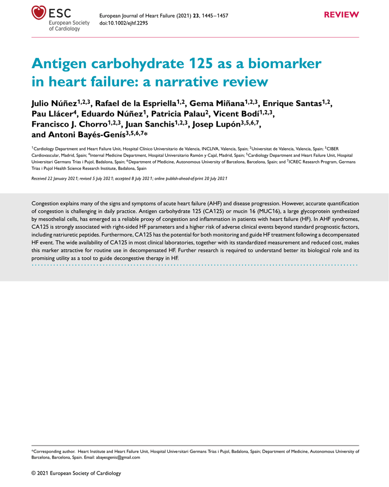
Antigen carbohydrate 125 as a biomarker in heart failure: a narrative review
Same clinical topic · Cardiology
Current ECG Aspects of Interatrial Block
Same clinical topic · Cardiology
Arrhythmic mitral valve prolapse in 2023: Evidence-based update
Same clinical topic · Cardiology
INTERPRETING THE NORMAL PACEMAKER
Same clinical topic · Cardiology